Modelos 3D do osso temporal – uma abordagem inovadora no treino cirúrgico otológico
DOI:
https://doi.org/10.34631/sporl.3124Palavras-chave:
Otologia, treino cirúrgico, impressão 3D, manufatura aditiva, modelos 3D, Osso temporal, simulação cirúrgica, planeamento cirúrgicoResumo
Introdução: A cirurgia do osso temporal exige elevada precisão, dada a complexidade da sua anatomia. O treino cirúrgico tradicional com cadáveres apresenta limitações como escassez de material, variabilidade anatómica e custos elevados. Este estudo explora a utilização de modelos 3D personalizados do osso temporal de doentes propostos para cirurgia otológica, como ferramenta inovadora de treino.
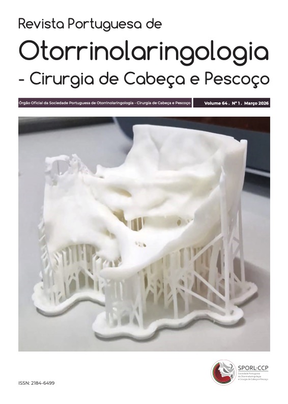
Material e Métodos: Foram criados modelos 3D personalizados a partir da TC de alta resolução do ouvido de cada doente e impressos em resina (White V4, impressora Formlabs 3+). O treino cirúrgico foi realizado antes da cirurgia in vivo. Posteriormente, os cirurgiões preencheram um questionário adaptado da Michigan Standard Simulation Experience Scale (MiSSES) para classificar a utilidade do modelo para treino cirúrgico.
Resultados: Foram utilizados oito modelos, abrangendo procedimentos como implante coclear, canalplastia e timpanomastoidectomia. Todos os inquiridos consideraram o treino útil, relatando maior confiança cirúrgica e potencial redução da taxa de complicações. Destacaram ainda o realismo da brocagem e a utilidade para treino de coordenação mão-olho.
Conclusão: Os modelos 3D do osso temporal representam uma ferramenta pedagógica promissora no treino cirúrgico otológico.
Downloads
Referências
Zagoog N, Yang VXD. State of robotic mastoidectomy: literature review. World Neurosurg. 2018 Aug;116:347-351. doi:10.1016/j.wneu.2018.05.194.
Hu M, Wattchow D, de Fontgalland D. From ancient to avant-garde: a review of traditional and modern multimodal approaches to surgical anatomy education. ANZ J Surg. 2018 Mar;88(3):146-151. doi:10.1111/ans.14189.
Rose AS, Webster CE, Harrysson OL, Formeister EJ, Rawal RB, Iseli CE. Pre-operative simulation of pediatric mastoid surgery with 3D-printed temporal bone models. Int J Pediatr Otorhinolaryngol. 2015 May;79(5):740-4. doi:10.1016/j.ijporl.2015.03.004.
Jiang Y, Jiang H, Yang Z, Li Y. The current application of 3D printing simulator in surgical training. Front Med (Lausanne). 2024 Aug 29:11:1443024. doi: 10.3389/fmed.2024.1443024.
Sun Z. Patient-specific 3D-printed models in pediatric congenital heart disease. Children (Basel). 2023 Feb 7;10(2):319. doi:10.3390/children10020319.
Saemann A, De Rosa A, Zubizarreta Oteiza J, Sharma N, Thieringer FM, Soleman J. et al. Innovating neurosurgical training: a comprehensive evaluation of a 3D-printed intraventricular neuroendoscopy simulator and systematic review of the literature. Front Surg. 2024 Nov 5:11:1446067. doi: 10.3389/fsurq.2024.1446067.
Yan M, Huang J, Ding M, Wang J, Song D. 3D-printed model is a useful addition in orthopedic resident education for the understanding of tibial plateau fractures. Sci Rep. 2024 Oct 22;14(1):24880. doi: 10.1038/s41598-024-76217-z.
Hunter SE, Freiberger JJ, Dear GdeL, Stolp BW, Moon R. A descriptive analysis of middle ear barotrauma in patients undergoing hyperbaric oxygen therapy. Undersea Hyperb Med. 2001;28(27).
Gurgel RK, Dogru S, Amdur RL, Monfared A. Facial nerve outcomes after surgery for large vestibular schwannomas: do surgical approach and extent of resection matter? Neurosurg Focus. 2012 Sep;33(3):E16. doi: 10.3171/2012.7.FOCUS12199.
Huang X, Xu J, Xu M, Chen M, Ji K, Ren J. et al. Functional outcome and complications after the microsurgical removal of giant vestibular schwannomas via the retrosigmoid approach: a retrospective review of 16-year experience in a single hospital. BMC Neurol. 2017 Jan 31;17(1):18. doi: 10.1186/s12883-017-0805-6.
Al Anazy FH, Alobaid FA, Alshiha WS. Sensorineural hearing loss following tympanoplasty surgery: a prospective cohort study. Egypt J Otolaryngol. 2016;32(2):93-97. doi:10.4103/1012-5574.181083
Gowrishankar SV, Fleet A, Tomasoni M, Durham R, Umeria R, Merchant SA. et al. The risk of meningitis after cochlear implantation: a systematic review and meta-analysis. Otolaryngol Head Neck Surg. 2023 Sep;169(3):467-481. doi: 10.1002/ohn.309.
Batsaikhan T, Seo YJ. Virtual reality simulators for temporal bone dissection: overcoming limitations of previous models. Res Vestib Sci. 2024;23(1):1-10. doi:10.21790/ rvs.2024.002.
Frithioff A, Frendo M, Pedersen DB, Sørensen MS, Wuyts Andersen SA. 3D-printed models for temporal bone surgical training: a systematic review. Otolaryngol Head Neck Surg. 2021 Nov;165(5):617-625. doi: 10.1177/0194599821993384.
Iannella G, Pace A, Mucchino A, Greco A, De Virgilio A, Lechien JR. et al. A new 3D-printed temporal bone: 'the SAPIENS'-specific anatomical printed-3D-model in education and new surgical simulations. Eur Arch Otorhinolaryngol. 2024 Sep;281(9):4617-4626. doi: 10.1007/s00405-024-08645-6.
Bento RF, Rocha BA, Freitas EL, Balsalobre FA. Otobone®: Three-dimensional printed temporal bone biomodel for simulation of surgical procedures. Int Arch Otorhinolaryngol. 2019 Oct;23(4):e451-e454. doi: 10.1055/s-0039-1688924.
Bartling M, Rohani SA, Ladak HM, Agrawal SK. Micro-CT of the human ossicular chain: Statistical shape modeling and implications for otologic surgery. J Anat. 2021 Oct;239(4):771-781. doi: 10.1111/joa.13457.
Molinari G, Emiliani N, Cercenelli L, Bortolani B, D'Azzero R, Burato A. et al. A novel 3D printed multi-material simulator for endoscopic stages surgery: the '3D Stages Trainer.' Laryngoscope. 2025 Apr 7. doi: 10.1002/lary.32168.
Frithioff A, Weiss K, Frendo M, Senn P, Mikkelsen PT, Sieber D. et al. 3D-printing a cost-effective model for mastoidectomy training. 3D Print Med. 2023 Apr 17;9(1):12. doi: 10.1186/s41205-023-00174-y.
Frithioff A, Weiss K, Senn P, Mikkelsen PT, Sørensen MS, Pedersen DB. et al. 3D-printed temporal bone models for training: does material transparency matter? Int J Pediatr Otorhinolaryngol. 2024 Sep;184:112059. doi: 10.1016/j.ijporl.2024.112059.
Bakhos D, Velut S, Robier A, Al zahrani M, Lescanne E. Three-dimensional modeling of the temporal bone for surgical training. Otol Neurotol. 2010 Feb;31(2):328-34. doi: 10.1097/MAO.0b013e3181c0e655.
Chien WW, da Cruz MJ, Francis HW. Validation of a 3D-printed human temporal bone model for otology surgical skill training. World J Otorhinolaryngol Head Neck Surg. 2021 Jan 14;7(2):88-93. doi: 10.1016/j.wjorl.2020.12.004.
Seagull FJ, Rooney DM. Filling a void: Developing a standard subjective assessment tool for surgical simulation through focused review of current practices. Surgery. 2014 Sep;156(3):718-22. doi: 10.1016/j.surg.2014.04.048.
Frithioff A. 3D-printed models for training temporal bone surgery [Thesis on the Internet]. [Copenhagen]: University of Copenhagen; Faculty of Health and Medical Science. 91 p. Available from: https://otonet.dk/AndreasFrithioff_thesis.pdf
Downloads
Publicado
Como Citar
Edição
Secção
Licença
Direitos de Autor (c) 2026 Sofia Sousa Teles, Ana Claro, Lília Ferraria, Mariana Neto, Rafael Pires, Leonor Oliveira, Joana Dias, Ana Miguel Couto, Ricardo Nelas, João Totxi, David Marcelo, Luís Antunes, Eduardo Simões, Rodolfo Peralta, Daniel Nunes

Este trabalho encontra-se publicado com a Licença Internacional Creative Commons Atribuição-CompartilhaIgual 4.0.